Human T -Cell Leukemia-Lymphoma Virus (HTLV): A Progress Report
 
R. C. Gallo, M. Popovic, P. Sarin, M. S. Reitz, Jr., V. S. Kalyanaraman, T. Aoki, M. G. Sarngadharan, and F. Wong-Staal    Hämatol. Bluttransf. Vol 28

A. Introduction

Since type-C retroviruses are known to be involved in naturally occurring leukemias of many animal species [26], a similar viral etiology has been sought in human leukemias. Some of the animal models provide important insight for consideration of human leukemias. For example, while most virus-induced animal leukemias and lymphomas are associated with abundant virus production in the tumor cells, bovine leukemia virus (EL V), the causative agent of bovine leukemias, was not detected until the leukemic cells were cultured in vitro (see review by Miller and Van Der Maaten [13]). This brings out the importance of long-term culture of the appropriate target cells for virus detection and isolation. In 1976, our laboratory reported the discovery of a factor termed T-cell growth factor (TCGF) [ 15]. Following interaction with an antigen, different subsets of mature T cells respond by making and releasing TCG F or making a receptor to TCG F. The TCG F binds to the receptor-bearing T cells and induces cell growth. Addition of exogenous TCGF can maintain growth of activated mature T cells for long periods [6, 26]. When TCGF was added to T cells obtained from patients with mature T -cell leukemias and lymphomas, some cells directly responded without prior activation in vitro [18]. Some of those samples released a retrovirus which we call human T -cell leukemia-Iymphoma virus (HTL V) ([19, 20]; 
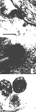
Popovic et al., in preparation). The morphology of HTL V is typically type c. Figure 1 shows an electron micrograph of some HTL V particles. Like all retroviruses, HTL V contains reverse transcriptase, has a high molecular weight RNA genome, and buds from cell membranes. It is distinct from all other known animal retroviruses [9, 16, 22, 23] and to date is the only unequivocal human retrovirus. (The retrovirus later isolated independently in Japan [14, 31] and called ATLV is, in fact, HTL V.) Furthermore, it is specifically associated with certain forms of human leukemia and lymphoma [4]. Here we wish to describe some of the new isolates of HTL V and report on some recent findings on the nature and distribution of HTL V and its transmission to and biological effects on normal T -lymphocytes in vitro.


B. Identification of New HTL V Isolates in Established Cell Lines
    Derived from Patients with T -Cell Leukemias/Lymphomas

Cell lines derived from patients with leukemia/lymphomas of mature T cells from geographically different parts of the world have been established in culture in the presence of TCGF as described previously for normal mature human T cells [ 15, 26] and neoplastic mature T cells [ 18 ]. These cell lines were analyzed for HTL V by (a) competition radioimmunoprecipitation assay (RIPA) for the major core protein p24 [9], (b) indirect immune fluorescence assay (IF A) using highly specific monoclonal antibody for another HTLV antigen, pl9 [24], (c) reverse transcriptase activity in the culture 
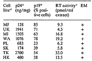
fluids, and (d) electron microscopy. In addition to the positive cell lines CR [19] and MB [20] published earlier, seven of eight recently established T -cell lines fully expressed HTL V (Table I) and one showed partial expression, These patients include four individuals from the United States, one from Israel, and three from a single Japanese family from the northwest part of the Honshu Island in Japan. In this family, the patient SK with acute T -cell lymphoma (A TL) and both his parents are virus positive. The father (MK) is clinically healthy and the mother has persistent lymphocytosis, which is considered to be a preleukemic state. All cell lines have karyotype and HLA patterns that match those of the donors, and the HLA profiles are different for all eight cell lines. In addition, a T -cell line established by Golde and colleagues from a patient (MO) with hairy cell leukemia [27] was positive vor HTL V p 19 and p24 [II]. We propose to designate each virus isolate with a subscript of the patient's initials, e.g., HTL V CR, HTL V MB, etc. All isolates except HTL V MO are highly related to each other as assayed by competitive radioimmunoassay with p24 (Fig. 2) and hybridization of viral cDNA to mRNA of the producer cell lines (Fig. 3). By these assays, the virus of Japanese A TL is indistinguishable from the prototype HTL V as exemplified by the earlier isolates HTL V CR and HTL V MB. On the other hand, HTL V MO competes poorly in the p24 assay (Fig.2), and nucleic acid sequence homology with HTL V CR was detected only under very nonstringent hybridization conditions ( our unpublished data). Therefore, this virus may form a distinct subgroup in the HTLV family. We propose to group them as HTLV-IcR, etc. versus HTLV-IIMo.


Fig. I. Electron microscopic examination of
the cellline MB, showing extracellular and
budding HTL V particles. Bar represents 90 µ


Table I. Expression of new HTL V isolates in T -cell lines
derived from patients with adult T -cellleukemia/lymphomas

a. Detected by competitive radioimmunoprecipitation
assay (RIPA) in cell extract

b. Indirect immunofluorescence assay (IFA)

c. Reverse transcriptase activity (RTA) in culture
fluids was measured with (dT)15(rA)n


d. Celllines were derived from peripheral blood (PE) or bone marrow (EM) of different patients as follows. (a) MJ from PB of a 50-yearold white male with mycosis fungoides, from Boston, Massachusetts; (b) UK from PB of a 45-year-old white male with diffuse histiocytic lymphoma, from Jerusalem, Israel; (c) MI from PB of a 32-year-old black female with T -cell lymphosarcoma cell leukemia, from Granada, West Indies; (d) WA from BM ofa 24-year-old black male with diffuse mixed lymphoma, from Augusta, Georgia; (e) PL from PB of a 27-year-old black female with T -cell diffuse mixed lymphoma, from Ovita, Florida; (I) SK from PE ofa 21-year-old male with adult T -cell leukemia, from Akita perlecture, Japan; (g) TK from PB of a 45-yearold female (mother of patient SK) who has 7% abnormal cells in her blood, from Akita perfecture, Japan; and (h) HK from PE of a 49year-old male (father of patient SK) who is normal, from Akita perfecture, Japan.


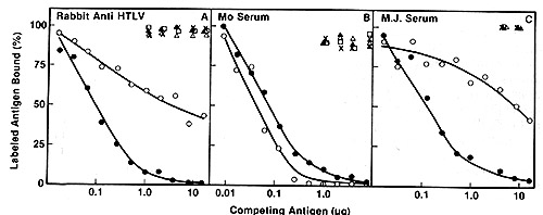
Fig.2A-C. Homologous and heterologous competition radioimmunoassays of HTL v p24. Assays were carried out as described [9] using l25I-labeled HTL V CR p24 and a limiting dilution of hyperimmune rabbit antibody to HTLV CR or sera from patients MO and MJ. A Competition RIA using rabbit anti-HTL V CR. B Competition RIA using MO serum. C Competition RIA using MJ serum. virus extracts used for competition were: black circle - black circle, HTLV CR; white circle- white circle , HTLV MO; x-x, Mason Pfizer monkey virus; white triangle- white triangle, bovine leukemia virus; white square - white square , Rauscher murine leukemia: black triangle - black triangle, simian sarcoma virus


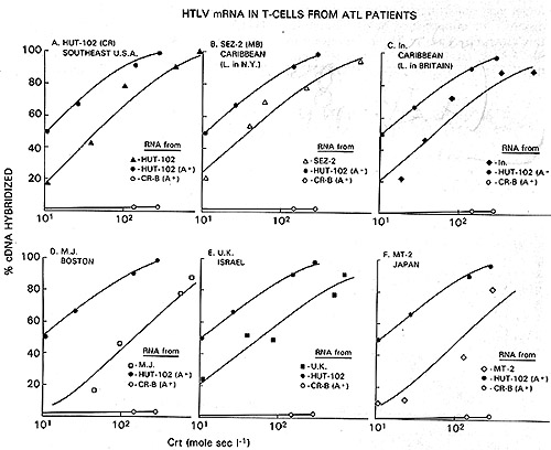
Fig.3A-F. Relationship of different HTL V isolates by RNA hybridization. ³H-HTL V (R cDNA was synthesized by calf thymus DNA primer [22] and hybridized to cellular RNA from different HTLVpositive cell lines. Results with the controls with poly(A) + RNA from CR- T (HUT 102) and CR-B cells are superimposed on each panel for comparison with the others


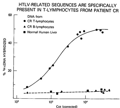
Fig.4. HTL V proviral DNA in T- but not B-celllines of patient CR, ³H-HTL V cDNA was annealed to cellular DNA from CR-T lymphocytes (black circle), CR-B lymphocytes (black triangle), and PHA-stimulated normal human peripheral blood T -lymphocytes (black square)


C. HTLV Provirus in Neoplastic T Cells: Evidence for Exogenous Infection

We had reported earlier that HTL V sequences are present in the infected cells and not in normal uninfected human cells [22], suggesting that HTL V is not an endogenous human virus. In the case of the patient CR, we also had the opportunity to find out whether he was infected pre- or post-zygotically [5]. Several T -cell lines, some clonal derivatives of these lines and a B-cellline have been established from CR. These cells were shown to have originated from the same individual by HLA typing. HTL V proviral DNA was detected in some but not all of the independently established T -cell lines of CR and not in the B cells. An example of the DNA hybridization kinetics is shown in Fig. 4. Furthermore, the surface phenotype OKT3-, OKT4+, and OKT8appears to correlate with the presence of HTL V. These results indicate that HTL V was acquired by CR by horizontal transmission and suggest that only a subtype of T cells is the target for HTL V infection.
Recently, molecularly cloned sequences representing the 5' and 3' ends of HTL V have been obtained in our laboratory [12 a]. These clones have been used as probes for Southern hybridization of fresh leukemic DNA from patients with HTL V -positive diseases [30]. These revealed one or few copies of HTL V integrated at a site which is unique for a given patient but varies from patient to patient. DNA from normal people did not contain hybridizing sequences. A similar observation has been made by others [31 ]. These results suggest that the infected cells are of clonal origin, so infection must have occurred prior to disease development. This feature is also found in animal leukemia-Iymphomas induced by chronic leukemia retroviruses.

D. Clinical Features of HTL V -Positive Diseases

Seroepidemiological studies have identified HTL V -positive patients from many regions of the world with at least three major areas that appear to be endemic: Southwestern Japan [4, 8, 9,25], the Caribbean [I], centraI South America (see also Blattner et al., this volume), but only sporadically in the United States [21 ]. Similar clinical features are found in the diseases associated with these areas, i.e., Japanese adult T -cellleukemia (ATL) and T-cell lymphosarcoma cell leukemia (T- LCL ) in the West Indian Blacks from the Caribbean. Both are represented by an aggressive course an frequent association with lymphadenopathy, hypercalcemia, hepatosplenomegaly, and cutaneous manifestations [2, 28]. The tumor cells are all mature, lack terminal deoxynucleotidyl transferase and express differentiated functions. Typing with monoclonal antibodies as well as functional studies showed that the cells may be either of the helper-inducer or suppressor-cytotoxic phenotype. Histologically, the cells are pleomorphic, often with highly convoluted nuclei. Almost all patients with A TL and T-LCL are HTLV positive. These observations led to the hypothesis that HTL V is associated with a subtype of adult T -cell malignancy which may include an aggressive form of cutaneous T -cell lymphoma (CTCL) found in patients CR and MB. In fact, the presence of HTL V may be of practical importance in disease classification. However, at least two HTL V -positive patients have relatively benign diseases: MJ with Sezary syndrome and MO with T -cell hairy cell leukemia. It should be noted, however, that at least the virus in MO is significantly 
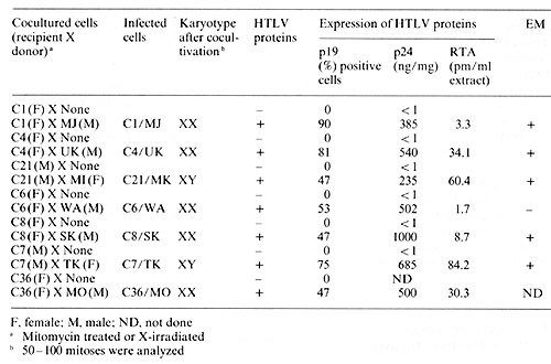
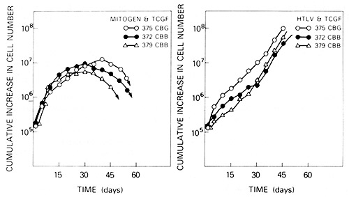
different from the prototype HTL V.

E. Infection and Transformation of Human Cord Blood T Cells by HTL V In Vitro

Seven of the HTL V isolates described above have been successfully transmitted into fresh human cord blood T cells by cocultivation (Popovic et al., in preparation). The virus-positive neoplasic cells used as donors were first treated with mitomycin-C or X-irradiation before cocultivation with recipient cord blood cells. After 4 weeks, assays for T -cell markers, HTL V, karyotype, and HLA-typing were performed. As shown in Table 2, all recipient cord blood are mature T cells, positive for HTL V provirus, and express various levels of HTL V antigens (pI9, p24, and RT). Karyotype and HLA typing consistently matched the recipient cells. Since cord blood T cells from the same donors were consistently negative for HTL V markers and the plasma from their cord blood were also negative for HTL V antibodies, we conclude that the virus was transmitted from HTL V -producing neoplastic T -cell lines into cord blood recipient T cells.
To characterize further whether a target for HTL V could represent a certain subset of mature T cells, phenotypes of HTL Vproducing cells were analyzed by a series of monoclonal antibodies specific for helper/ inducer and suppressor/cytotoxic T cells. We found that a majority of HTL V -producing T -cell lines consistently exhibited only helper-inducer phenotype. Two established T -cell lines, SK and TK, both from Japanese patients and two HTL V -infected cord blood T cells (C 1 and C5) revealed "double" phenotype. However, none of the T -cell lines exhibited pure suppressor/cytotoxic phenotype. Unlike HTL V-infected cord blood T cells, PHAstimulated cells (control) consist of 70% helper/inducer and 30% of T cells with suppressor/cytotoxic phenotype. Thus, these data from T -cell phenotype characterization of HTL V -infected T cells again suggest that a certain subset of mature T -cells is the target for HTL V. HTL V infection studies with cord blood cells deprived ofT-cell population with helper/ind ucer of suppressor / cytotoxic phenotype are currently being carried out.
HTL V -infected cord blood T cells differ from mitogen-stimulated cord blood T cells in several growth properties and cell surface characteristics, the infected cells resembling more the neoplastic cells transformed in vivo by HTL V (see Sarin et al., this volume for details). The most striking feature of HTL V -infected cord blood T cells is their potential for indefinite growth as shown in Fig.5. In contrast, mitogenstimulated cord blood T cells from the same patients consistently exhibited growth "crises" after 1 month in culture, even in the continued presence ofTCGF. Furthermore, the infected cells, like the neoplastic cells, had the tendency to form clumps in culture. When analyzed by electron microscopy, the cells were seen to have convoluted nuclei (not shown) while the mitogen-stimulated cells did not. Another important and reproducible difference is the decrease in requirement for TCGF by the infected cells. In fact, some of the infected cells are completely independent of exogenous TCGF (see Sarin et al., this volume). Other changes of the infected cells include alteration in their HLA profile and expression of receptors for transferrin, TCGF, and HAA (human-activated lymphocyte antigen detected by monoclonal antibodies) in a high percentage of cells. The data indicate that HTL V is also capable of causing morphological transformation of cord blood T cells in vitro (see Sarin et al., this volume).

Table 2. Transmission of HTLV into human cord blood T cells


Fig.5. Growth curves of uninfected and HTL V -infected human cord blood T cells in vitro. Left panel, mitogen-stimulated cord blood T cells. Right panel, HTL V -infected cord blood T cells. C6/W A and C7/TK cell lines are primary cocultures (for details see Table 2). C5/MJ cells were obtained in three successive transmissions of HTL V MJ isolate into cord blood cells

F. Possible Molecular Mechanism of Transformation by HTL V

As mentioned earlier, analysis of HTL V positive leukemic T cells showed that the cells are of clonal origin with respect to the provirus integration sites. In animal systems monoclonality has also been shown to be a common feature of leukemias induced by retroviruses which are chronic leukemia viruses but not those induced by retroviruses which are acute leukemia viruses. Consequently, in spite of its high efficiency to transform T cells in vitro, HTL V probably does not carryon onc gene. Several chronic leukemia viruses are known to induce leukemia by activating cellular onc genes (myc in B-cell lymphomas and erb in erythroleukemias) ([7]; Kung, personal communication) by integrating in the proximity of these genes. Activation of thcse genes is brought about by providing either a viral promotor or viral nucleotide sequences dubbed ""enhancer" [12, 17], the real function of which is still unknown. Since HTL V specifically transforms mature T cells, it is likely to affect expression of genes that are important in T -cell proliferation. A model has been proposed for the mechanism of leukemogenesis by HTL V [4]. Briefly, the HTL V envelope protein interacts with the population ofT cells normally designed to make TCGF receptors, mimicking an antigen stimulation of blastogenesis. These cells then synthesize receptors for TCGF. Simultaneously, the HTLV provirus integrates in the vicinity of the TCGF gene or a gene that exerts a pleiotropic effect on TCGF expression and activates this gene either hy direct promotion or enhancement. The production ofTCGF by a cell bearing a TCGF receptor may result in autostimulation and increased cell proliferation. As an approach to study the gene(s) activated by HTL V infection, we have recently identified and isolated a gene that is expressed at high levels in all HTL V -positive neoplastic T cells and in normal cord blood T cells after infection with HTLV hut not the uninfected counterparts [12 b]. Study of the expression pattern of this gene in uninfcctcd human hematopoietic cells suggests that its expression may be linked to TCGF production. Experiments are in progress to determine if HTL V integrates at a preferred locus in the human chromosome and affects transcription of specific cellular genes in the vicinity, including this gene in question.

References

1. Blattner WA, Kalyanaraman VS, RobertGuroff M, Lister TA, Galton DAG, Sarin PS, Crawford MH, Catovsky D, Greaves M, Gallo RC (1982) The human type-C retrovirus, HTL V, in Blacks from the Caribbean region, and relationship to adult T -cell leukemia/lymphoma. Int J Cancer 30:225-264

2. Catovsky D, Greaves MF, Rose M, Galton DAG, Goolden AWG, McCluskey DR,White JM, Lampert I, Bourikas G, Ireland R, Brownell AI, Bridges JM, Blattner W A, Gallo RC (1982) Adult T -cell Iymphomaleukaemia in blacks from the West Indies. Lancet 1:639-642

3. Gallo RC (1982) Regulation of human T-cell proliferation. T-cell growth factor, T-cell leukemias and lymphomas, and isolation of a new C-type retrovirus. In: Rosenberg SA, Kaplan HS (eds) Malignant lymphomas. Academic, New York, pp 201-218

4. Gallo RC et al. (to be published) The human type-C retrovirus. association with a subset of adult T -cell malignancies. (Submitted)

5. Gallo RC, Mann D, Broder S, Ruscetti FW, Maeda M, Kalyanaraman VS, RobertGuroffM, Reitz Jr MS (1982) Human T-cell leukemia-Iymphoma virus (HTLV) is in T but not B lymphocytes from a patient with cutaneous T -cell lymphoma. Proc Nat Acad Sci USA 79.5680-5683

6. Gootenberg JE, Ruscetti FW, Mier JW, Gasdar A, Gallo RC (1981) Human cutaneous T -cell lymphoma and leukemia cell lines produce and respond to T -cell growth factor. JExp Med 154.1403-1418

7. Hayward WS, Neel BG, Astrin SM (1981) Induction of lymphoid leukosis by avian leukosis virus: activation of a cellular "onc" gene by promoter insertion. Nature 290:475-480

8. Hinuma Y, Nagata K, Hanaoka M, Dakai M, Matsumoto T, Kimoshita KI, Shivakawa S, Miyoshi I ( 1981 ) Adult T -cell leukemia: antigen in an A TL cell line and detection of antibodies to the antigen in human sera. Proc Nat Acad Sci USA 78:6476-6480

9. Kalyanaraman VS, Sarngadharan MG, Poiesz BJ, Ruscetti FW, Gallo RC (1981) Immunological properties of a type C retrovirus isolated from cultured human T -lymphoma cells and comparison to other mammalian retroviruses. J Virol 38. 906-913

10. Kalynaraman VS, Sarngadharan MG, Nakao Y, Ito Y, Aoki T, Gallo RC (1982) Natural antibodies to the sturctural core protein (p24) of the human T-cell leukemia (lymphoma) retrovirus (HTL V) found in sera of leukemic patients in Japan. Proc Nat Acad Sci USA 79.1653-1657

11. Kalyanaraman VS, Sarngadharan M G , Robert-Guroff M, Miyoshi 10, Blayney D, Golde D, Gallo RC (1982) A new subtype of human T-cell leukemia/lymphoma virus (HTL V -II) associated with a T -cell variant of hairy cell leukemia. Science 218:571-573

12. Levinson B, Khoury G, Vande Woude G, Gruss P (1982) Activation of SV40 genome by 72 base pair tandem repeats by Moloney sarcoma virus. Nature 295: 568-572

12a. Manzari V, Wong-Staal F, Franchini G, Colombini S, Gelmann EP, Oroszlan S, Staal SP, Gallo RC (to be published) Human T -cell leukemia-Iymphoma virus, HTL V. molecular cloning of an integrated defective provirus and flancing cellular sequences. Proc Nat Acad Sci USA

12b. Manzari V, Gallo RC, Franchini G, Westin E, Ceccherini-Nelli L, Popovic M, WongStaal F (to be published) Abundant transcription of a cellular gene in T -cells infected with human T -cell leukemia-Iymphoma virus (HTLV). Proc Nat Acad Sci USA

13. Miller JM, Van Der Maaten MJ (1980) The biology of bovine leukemia virus infection in cattle in viruses. In. Essex M, Todaro G, zur Hausen H (eds) Naturally occurring cancers. Cold Spring Harbor conferences on cell proliferation vol. 7. Cold Spring Harbor Press, New York, pp 901-910

14. Miyoshi I, Kabonishi I, Yoshimoto S, Akagi T, Ohtsuki Y, Shiraishi Y, Nagata K, Hinuma Y ( 1981 ) Type C virus particlcs in a cord T -cell line derived by co-cultivating norma] human cord leukocytes and human leukemic T -cells. Nature 294: 770- 771

15. Morgan DA, Ruscetti FW, Gallo RC (1976) Selective in vitro growth of T -lymphocytes from normal human bone marrows. Science 193:1007-1008

16. Oroszlan S, Sarngadharan MG, Copeland TD, Kalyanaraman VS, Gilden RV, Gallo RC (1982) Primary structure analysis of the major internal protein p24 of human type C T -cell leukemia virus. Proc N at Acad Sci USA 79: 1291-1294

17. Payne GS, Courtneidge SA, Crittenden LB, Fadly AM, Bishop JM, Varmus HE (1981) Analysis of avian leukosis virus DNA and RNA in bursal tumors: viral gene expression is not required for maintenance of the tumor state. Cell 23:311-322

18. Poiesz BJ, Ruscetti FW, Mier JW, Woods AM, Gallo RC (1980a) T-cell lines established from human T -lymphocytic neoplasias by direct response to T -cell growth factor. Proc Nat Acad Sci USA 77:6815-6819

19. Poiesz BJ, Ruscetti FW, Gazdar AF, Bunn PA, Minna JD, Gallo RC (1980b) Isolation of type-C retrovirus particles from cultured and fresh lymphocytes of a paticnt with cutaneous T-cell lymphoma. Proc Nat Acad Sci USA 77:7415-7419

20. Poiesz BJ, Ruscetti FW, Rcitz MS, Kalyanaraman VS, Gallo RC (1981) Isolation of anew type C retrovirus (HTL V) in primary uncultured cells of a patient with Sezary T -cell leukaemia. Nature 294.268-271

21. Posner LE, Robert-Guroff M, Kalyanara man VS, Poiesz BJ, Ruscetti FW, Fossieck B,Bunn PA, Minna JD, Gallo RC (1981) Natural antibodies to the human T -cell lymphoma virus in patients with cutaneous T -celllymphomas. J Exp Med 154.333-346

22. Reitz MS, Poiesz BJ, Ruscetti FW, Gallo RC ( 198 I) Characterization and distribution of nucleic acid sequences of a novel type C retrovirus isolated from neoplastic human T lymphocytes. Proc Nat Acad Sci USA 78: 1887-1891

23 Rho HM, Poiesz BJ, Ruscetti FW, Gallo RC ( 1981) Characterization of the reverse transcriptase from a new retrovirus (HTL V) produced by a human cutaneous T -cell lymphoma cell line. Virology 112:355-358

24. Robert-Guroff M, Ruscetti F, Posner LE, Poiesz BJ, Gallo RC (1981) Detection of the human T -cell lymphoma virus p 19 in cells of some patients with cutaneous T -cell lymphoma and leukemia using a monoclonal antibody. J Exp Med 154: 1957-1964

25. Robert-Guroff M, Nakao Y, Notake K, Ito Y, Sliski A, Gallo RC (1982) Natural antibodies to human retrovirus HTL V in a cluster of Japanese patients with adult T -cell leukemia. Science 215:975-978

26. Ruscetti FW, Gallo RC (1981) Human T -lymphocyte growth factor: the second signal in the immune response. Blood 57. 379-393

27 Saxon A, Stevens RH, Golde DW (1978) T -lymphocyte variant of hairy cell leukemia. Ann Int Med 88.323-326

28. Uchiyama T, Yodoi J, Sagawa K, Takatsuki K, Uchino H ( 1977) Adult T -cell leukemia. clinical and hematological features of 16 cases. Blood 50.481-503

29. Wong-Staal F, Gallo RC (to be published) Retroviruses and leukemia. In: Gunz F, Henderson E (eds) Leukemia. Grune and Stratton, New York

30. Wong-Staal F, Hahn B, Manzari V, Colombini S, Franchini G, Gelmann EP, Gallo RC (to be published) A survey of human leukemias for sequences of a human retrovirus, HTLV Nature

31. Yoshida M, Miyoshi I, Hinuma Y (1982) Isolation and characterization of retrovirus from cell lines of human adult T -cell leukemia and its implication in the disease. Proc Nat Acad Sci USA 79: 2031-2035

Read more

  • "RNA Tumor Viruses and Leukemia: Evaluation of Present Results Supporting their Presence in Human Leukemias", 1976
  • "Cellular and Virological Studies Directed to the Pathogenesis of the Human Myelogenous Leukemias", 1979
  • Personal Reflections on the Origin of Human Leukemia 1987
  • Human Retroviruses: Linkage to Leukemia and AIDS 1992