|
1 Institut für Virologie der FU Berlin
2 Robert-Koch-lnstitut des Bundesgesundheitsamtes
3 Institute of Cancer Research, Chester Beatty Laboratories, London
4 Deutsches Primatenzentrum, Göttingen
5 National Cancer Institute, Bethesda, MD
6 New England Regional Primate Res. Center, Southborough
LA V /HTL V -III was investigated by thinsection and immunoelectron
microscopy. Formation of the virion takes place at the cell membrane.
The inner components are assembled concomitant with budding, as
is characteristic of type-C oncovirinae. Different from type-C viruses
and typical for the subgroup of lentivirinae, these components in
immature particles form an 18-nm broad, electron-dense spherical
shell apposed to the viral membrane. After budding the structural
components are rearranged ("maturation"). The electron-dense nucleoid
formed is surrounded by a prismatic electronopaque core shell 4-5
nm thick [1]. From immunocryoultramicrotomy, using monoclonal as
well as monospecific antibodies, we concluded that the core shell
is built up by p24. The inner leaflet of the envelope is covered
by a 5-7-nm electron-dense layer with p18 antigenicity. Adjacent
to this layer we observed electron-dense "lateral bodies" with unknown
composition and function. Knobs on the viral envelope can be demonstrated
with tannic acid-treated samples. While on budding or immature particles
a uniform fringe of equidistantly spaced spikes is visible, which
can be labelled with anti-gp120 and/ or anti-gp41 antibodies, mature
virions lack these projections partially or completely. The spontaneous
loss of knobs seems to be rapid. This became particularly evident
when parallel cultures harvested at different times were investigated.
Five- to 7-day-old cultures only rarely contained spiked particles.
The knobs, about 70-80 per virion, have a height of 9 nm above the
unit membrane and a diameter of 15 nm. They are connected to the
virion via stalks 7-8 nm thick [2] .
When patient sera were investigated by IEM it could be shown that
LA V /HTL V III-infected individuals carry antibodies directed against
different viral proteins. IEM revealed a qualitative correlation
of labelling intensity of viral envelope components and neutralizing
capacity. The presence of antibodies in patients and the assumption
that envelope proteins are also shed in vivo have important implications.
First, protecting antibodies might be captured by shed proteins
and are therefore not available for neutralization. Second, circulating
immune complexes (CIC) can be involved in the pathogenicity of LAV/HTLV-III.
CIC of unknown composition have been frequently observed in ARC
and AIDS patients. An improvement of clinical symptoms after plasmapheresis
of CIC has been reported [3]. The question whether LA V /HTL V -III
antigens are involved in the formation of CIC can be answered by
the characterization of such complexes.
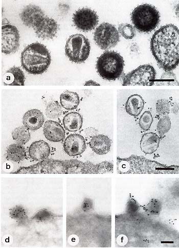
Fig. 1. a Ultrathin sections of LA V /HTL V III particles after
treatment with tannic acid and Epon embedding. "Immature" particles
just after budding are densely studded with knobs. The viral RNP
is closely apposed to the viral membrane as a concentric shell.
"Mature" particles lack the fringe of projections and show an elongated
tubular core and ill-defined "lateral bodies". b, c Pre embedding
IEM of LA V /HTL V III using antigp120 (b) or anti-gp41 (c) peptide
antisera. d, e Immuno-gold labelling of ultrathin cryosections after
incubation with a p24-specific monoclonal antibody (d) and a mono
specific anti-p24 antibody (e). f Labelling after incubation with
antip18 hyperimmune serum leads to a shell-Iike distribution of
the marker.
References
1. Gelderblom H, Özel M, Pauli G (1985) T -Zellspezifische Retroviren
des Menschen: V erglei chende morphologische Klassifizierung und
mögliche funktionelle Aspekte. Bundesgesundhbl.28:161-171
2. Gelderblom HR, Hausmann EHS, Özel M, Pauli G, Koch MA (1987)
Fine structure of human immunodeficiency virus (HIV) and immunolocalization
of structural proteins. Virology 156:171-176
3. Kiprov DD, Lippert R, Sandstrom E, Jones FR, Cohen RJ, Abrams
D, Busch DF (1985) Acquired immunodeficiency syndrom (AIDS)-apheris
and operative risk. J Clin Apheresis 2:427-440
|