|
* Supported by grants CA 20194, CA 32516 and KO8-CA
00966 (JG) from the National Cancer Institute, A. Introduction Within the family of hematopoietic growth factors, murine G-CSF was first recognised as acting predominantly upon mature neutrophils and neutrophil progenitors, stimulating murine neutrophil colonies characterized by small size, maturity, and relative infrequency [1, 2]. G-CSF is synthesized in many murine tissues and organs, possibly by common populations ofmacrophages or endothelial cells [3], and the factor has been purified from the lung tissue of endotoxintreated mice as a 24000-25000 Mr glycoprotein [4]. In contrast to GM-CSF and interleukin-3 (IL-3), G-CSF exerts a potent differentiation-inducing action on myeloid leukemic cells such as the WEHI-3 myelomonocytic leukemic cell line [ 4-6] and the M 1 myeloblastic leukemic line [7]. Characterization of human CSF in human placental [8] and bladder carcinoma cell line [9] conditioned medium by hydrophobic chromatography or by ion-exchange chromatography and reversed phase-high performance liquid chromatography (RP-HPLC) [10, 11] revealed two species. The least hydrophobic activity, called CSF -alpha [8, 9] or pluripoietin alpha [11], is structurally and functionally homologous to murine GM-CSF (al though not species cross-reactive), and the hydrophobic species called CSF-beta [8, 9] or pluripoietin [2, 10] is the human analogue of murine G-CSF since these two CSFs exhibit similar activities on murine and human cells [2, 8, 10] and are fully cross-reactive with each other's specific cellular receptors [8]. This latter CSF species was purified to homogeneity from the conditioned medium of the human bladder carcinoma cell line 5637 and \\'as shown to be O-glycosylated and to have a molecular weight of 19600 [10]. The gene encoding this pluripotent human G-CSF was subsequently cloned and expressed in E. coli [12]. The E. coli rhGCSF was comprised of 174 amino acids with a deduced molecular weight of 18700 and it had no significant homology with any other previously sequenced growth factors. Recently, the cDNA sequence coding for murine G-CSF has been isolated from a cDNA library prepared with mRNA derived from murine fibrosarcoma NFSA cells, which produce G-CSF constitutively [13]. The nucleotide sequence and the deduced amino acid sequence of murine G-CSF cDNA were 69% and 73% homologous, respectively, to the corresponding sequences of human GCSF cDNA. Native and recombinant G-CSF had comparable biological activity, stimulating neutrophil granulocyte colonies of mouse and man with a specific activity of 1 x 10⁸ units/ mg of pure protein [12]. Our earlier observations with purified G-CSF, showing that at high concentrations the factor stimulated BFU-E and CFU-GEMM in methylcellulose cultures of adherent and T -cell-depleted human marrow [10], were confirmed using rhG-CSF [12]. More recent studies in our laboratory [14], using highly enriched hematopoietic progenitor cells, failed to demonstrate a direct effect of human G-CSF on BFU-E and CFU-GEMM, suggesting that the earlier observations may have been the consequence of an indirect mechanism involving non- T accessory cells. In this context, sequential observations in mouse bone marrow culture indicate that murine G-CSF can initiate proliferation in many progenitor cell populations, including multipotential and erythroid progenitors, but fails to sustain proliferation in these lineages beyond 24 days [15]. The action of G-CSF on mature hematopoietic cells appears to be confined to an action on neutrophils involving increased expression of chemotactic receptors, enhanced phagocytic ability, cellular metabolism associated with the respiratory burst, antibody-dependent cell killing, and the expression of function-associated cell surface antigens [2,16]. The action of G-CSF on leu kemic cells is receptor mediated, and competitive binding studies with 1251-labeled hG-C SF [12] or murine G-CSF [9] revealed receptors on fresh human leukemic marrow cells classified as M2, M3, and M4 and on murine WEHI-3 and human HL-60 and U937leukemic cell lines. Furthermore, these leukemic cell lines and receptor-positive human leukemic cells were induced by recombinant G-CSF to undergo terminal differentiation to macrophages and granulocytes [12]. The ability of endotoxin to elicit production of G-CSF can account for most reports of the leukemia-differentiating action of postendotoxin serum in mouse and man [6, 17]. However, the elevation of bioactive G-CSF in postendotoxin serum is partially masked at high serum concentrations by an inhibitory activity which is particularly evident in postendotoxin sera of mice primed with bacille Calmette-Guerin (BCG) or C. parvum [6,17]. This hematopoietic or colony inhibitory activity was shown to be directed at both normal CFU -0 M and myeloid leukemic cells, and biochemical characterization showed that it copurified with an activity with both in vitro L-cell cytotoxicity and in vivo tumor necrosis action [6, 17]. Subsequent studies distinguished between the antiproliferative effects of tumor necrosis factor (TNF) on CFU-OM and myeloid leukemic cells and the proliferation- and differentiation-inducing action of the G-CSF coinduced with similar kinetics in the serum of C. parvum-endotoxin-treated mice [17]. With the availability of recombinant human TNF alpha [18], it was possible to demonstrate that human CFU-GM, BFU-E,and CFU-GEMM were inhibited by relatively low concentrations of TNF ( < 100 units/mI) and that the inhibition was potentiated by the addition of gamma interferon [19]. We have explored the antagonism between CSF species and TNF alpha in both short-term clonogenic assays and long-term bone marrow cultures and have obtained evidence that TNF may playa physiological role in antagonising G-CSF action in generation of neutrophil granulocytes.
For human assay, 2.5 x 10 high 4-1 x 10 high 5 lowdensity, nonadherent, and T -cell-depleted normal marrow or unseparated cells from long-term bone marrow culture (L TBMC) were cultured in 0.3% agar in 1 ml supplemented McCoy's medium in the presence of 200-1000 units rhG-CSF or rhGM-CSF. In some studies, 5% 5637 cell line CM, or G- or GM-CSF purified from this source were used as sources of stimuli. Colonies were scored at days 7 and 14. For murine assay, 2.5 x 10 high 4 marrow cells or 1 x 10⁵ spleen cells were cultured in Iscove's Modified Dulbecco's medium containing 15% fetal calf serum (FCS) and 0.3% agarose. Cultures were stimulated with interleukin-3 (IL-3) purified from WEHI-3 CM, CSF-1 purified from L-cell CM, G M -CSF purified from mouse lung CM, and rhO-CSF at 200-1000 units/mI. Cultures were scored at 5- 7 days and in some studies, colony morphology was determined on fixed, stained agar plate preparations.
The differentiation-inducible D + sub line of the WEHI-3B murine myelomonocytic leu kemic cell line was used to monitor the differentiation-inducing action of G-CSF [6, 10]. 3 x 10˛ WEHI-3 cells were incubated in 0.3% agar in supplemented McCoy's medium containing 12.5% FCS with 0.2 ml/ well in 24-well tissue culture trays (Costar, Cambridge, MA) and in replicates at 37 °C in 5% CO2 in air. Cultures were scored on day 7 for induction of dispersed, differentiated colonies or tight, blast-cell colonies. Total cloning efficiency was close to 30% at 7 days of culture and was not changed significantly in the presence of G-CSF .
Obtained by aspiration from the posterior iliac crest of healthy volunteers who had given informed consent, 107 normal human bone marrow nucleated cells were inoculated into 25 cm tissue culture flasks in 10 ml McCoy's medium containing 15% FCS, 15% horse serum, and 10high -6 M hydrocortisone ("Gartner's medium"). After being gassed with 5% CO2 in air, the cultures were incubated at 37 °C for 3 days, all suspension cells removed and Ficoll-Hypaque (Pharmacia) separated, and were returned to the culture flasks, together with fresh medium and the appropriate concentration of rhTNF-alpha (a generous gift from Cetus, Inc.) or rhG-CSF (provided by Amgen). Long-term cultures were subject to demidepopulation of suspension cells and medium at weekly intervals and assayed for total cells, differential count, and CFU-GM at days 7 and 14 in agar assays stimulated with G- or GMCSF.
C3H/HeJ mice, 8-12 weeks old, purchased from Jackson Laboratories and maintained in our Institute in laminar air-flow rooms were injected intraperitoneally twice daily with 1.5 µg rhG-CSF in 0.2 ml hydroxethylpiperazine ethanesulphonic acid (HEPES) buffered balanced salt solution (BSS) containing 1.5 µg purified bovine serum albumin (BSA). Control inocula consisted of buffer containing 3 µg BSA and 1.5 ng E. coli endotoxin (Difco ). The endotoxin contamination of the rhG-CSF was < 0.5 µg/ mg of protein. Mice were bled retro-orbitally for total white blood cell count (WBC) and hematocrit, and a smear was obtained for a blood differential. After 1 or 2 weeks of G-CSF treatment, groups of three mice were killed and cell suspensions prepared from femurs, spleen, and thymus. In addition, peritoneallavage with 10 ml ice-cold BSS containing 10 units/mI heparin was used to obtain unstimulated peritoneal exudate suspensions. Liver hematopoietic cell populations were obtained by collagenase treatment of a minced liver preparation, with subsequent passage through a wire mesh sieve and percoll separation to remove debris and hepatocytes. Cytocentrifuge preparations of lymphohematopoietic tissues were made for morphological assessment, and cell suspensions were assayed for CFU-GM, BFU-E and CFU-GM in standard clonogenic assays. In addition, hematopoietic (CFU-S) determinations were made by injecting 10 high 5 cells intravenously into groups of 5 C3H/HeJ mice lethally irradiated with 950 rad gamma irradiation. Mice were killed after 12 days and colonies enumerated on Bouin's-fixed spleens.
I. Interaction Between G-CSF, GM-CSF and TNF Alpha on Human Myeloid Colony Formation As previously reported [11 ], colonies stimulated by GM-CSF were
maximal at day 14 of culture, when approximately one third of colonies
contained eosinophils and the remainder were neutrophil or mixed
neutrophil-macrophage, and at day 7 most clones were of subcolony
size «40 cells). In contrast, with rhG-CSF, neutrophil clones of
colony size were present at day 7 and their numbers remained approximately
constant to day 14, when the majority were neutrophil and a minority
( < 20% ) were neutrophil-macrophage mixed. Synergism between G-CSF
and GM-CSF was noted in the context of increased colony size and
number. Dose-response analysis revealed that a more 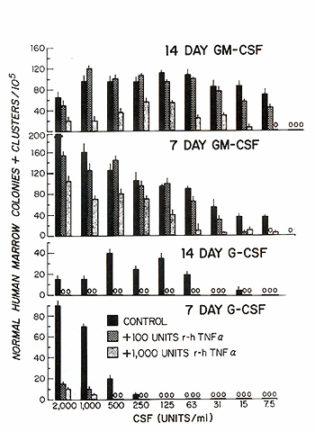
shallow titration of response was evident when day 14 was the endpoint, and at both days 7 and 14 the G-CSF dose-response curve was steeper than that of GM-CSF when either colonies or colonies plus clusters were used as the endpoint (Fig. 1). The inhibitory influence of rhTNF alpha on the CFU-GM assay has been reported elsewhere [19], but we have observed additional variables that determine the degree of inhibition, specifically the quantity and species of CSF used to stimulate the assay and the influence of accessory cell populations. With unseparated normal human marrow, colony formation was not significantly inhibited by 100 units/mI TNF when cultures were stimulated with 30-2000 units GMCSF, and with 1000 units TNF 20%-80% inhibition was seen; in both instances, comparable inhibition was seen at days 7 and 14 of culture (Fig. 1 ). With G-CSF as a source of stimulus, > 80% inhibition of colony and cluster formation was seen at day 7 with 100 units TNF even in the presence of high concentrations of stimulus, and at day 14 total inhibition of colony formation was ob served. Particularly evident in the case of GCSF at concentrations of < 500 units and to a lesser extent with GM-CSF at < 30 units, the degree of TNF inhibition was amplified at these lower, nonplateau levels of stimulation. Removal of mature granulocytes and monocyte-macrophage populations by adherence and density separation did not significantly alter the TNF dose-response curve since 50% day-14 colony inhibition was seen with 50 units rhTNF in cultures stimulated with 1000 units G-CSF and 400 units TNF was required to give comparable inhibition in cultures stimulated with 1000 units GMCSF (Table 1). Additionally, while all colony formation was inhibited in G-CSFstimulated cultures with 500 units TNF, a substantial number of colonies (22% ) were found to be resistant to TNF even at concentrations of 10000 units in GM-CSF-stimulated cultures. Further depletion of accessory cells and enrichment for progenitor cell populations using immunoadherence ""panning" and complement-mediated cytotoxicity increased the sensitivity of G-CSF -stimulated CFU-GM to TNF inhibition but did not alter the sensitivity of the GM-CSFstimulated colonies (Table 1). Fifty percent inhibition of G-CSF -stimulated colonies in such accessory cell-depleted marrow was now evident with 5 units TNF, a 2-log lower concentration than required to produce comparable inhibition in GM-CSF-stimulated cultures. Table I. Inhibitory activity of TNF on
human CFU-GM stimulated by G or GMCSF 
II. Action of G-CSF and TNF in Human Long-term Bone Marrow Culture The inoculation of 107 normal human marrow cells in 10 ml of Gartner's
medium, with weekly demidepopulation and no additional recharging,
resulted in the establishment of a confluent, adherent layer of
marrow stromal cells with extensive adipocyte development and sustained
hematopoiesis. After an initial falloff from the maximum cellularity
in the 1 st week, total cells were produced at a level of 7.4 x
105 per flask for the first 2 months with polymorphonuclear neutro
phils (PMN) comprising 12%-52% of total cells (mean of 35%, Fig.2).
CFU-GM responsive to GM-CSF fell from a maximum of 29000 per flask
at 1 week to 1600 after 2 months, and G-CSF-responsive CFU-GM were
28800 at 1 week and 2200 at 2 months. Neutrophil and CFU-GM production
persisted for 14 weeks. The weekly addition of rhG-CSF (10000 units/mI)
resulted in a marked, sustained elevation of neutrophil production.
Between 2 and 8 weeks of culture, cell production averaged 4.0 x
106 neutrophils per flask, a level of neutrophil production ten
times higher than in control cultures, sustained for 2 months. CFU-GM
production in the early stages of culture in the presence of G-CSF
exceeded control values by approximately a factor of 2 regardless
of whether the colonies were stimulated by G- or GM-CSF, and no
evidence was found for depletion or premature exhaustion of G-CSF-stimulated
long-term marrow cultures, since despite the chronic elevation of
mature neutrophil production, total CFU-GM produced by 2 months
were comparable in control and G-CSF-stimulated culture. In contrast
to the proliferative stimulus provided by G-CSF, TNF inhibited neutrophil
production and CFU-GM production. At concentrations of 1000 units/mI
TNF alpha, cell production was 50% of control in the first 3 weeks,
25% by 4 weeks and only 7% of control with no neutrophil production
by 9 weeks; no CFU-GM were produced after 5-6 weeks. At lOO units/mI
TNF no difference from control cultures was evident ti118-9 weeks,
but by 9 weeks cellularity was 7.5% of control and PMN production
was 2% of control with no CFU-GM produced. The lowest concentration
of TNF tested (10 units/mI) produced an average 50% reduction in
PMN production, otherwise no difference in either CFU-GM production
or culture longevity was found. These observations were reproduced
in four subsequent experiments and in one study ( data not shown),
attempts to reverse the TNF -mediated suppression of myelopoiesis
by coaddition of G-CSF were unsuccessful. 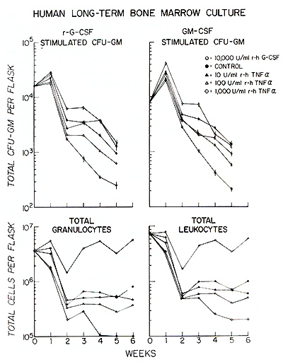
The preceding studies on G-CSF action in long-term marrow culture indicated that in the first few weeks of culture the total number of CFU-GM responsive to either GCSF or GM-CSF was increased substantially over control values. This observation, together with earlier studies indicating that G-CSF could generate CFU-GM in simple 1-week suspension cultures of human mar row in a "pre-CFU-GM assay" [2], suggested that G-CSF acted on a cell earlier in the developmental lineage than the CFU GM or alternatively promoted enhanced CFU-GM self-renewal. A third explanation could be that some accessory cell population produced a factor in response to G-CSF that caused recruitment of new CFU-GM by an action on a precursor cell population. In our earlier studies using the "pre-CFU-GM" assay, adherence and T -cell depletion were used to remove some types of accessory cell; however, it is known that other hematopoietic growth factor-producing cells are not removed by this procedure, e.g., natural killer (NK) cells. We therefore subjected normal human marrow to a more rigorous accessory and differentiating cell depletion procedure using a spectrum of monoclonal antibodies in conjunction with immunoadherence panning and complement-mediated cytotoxicity [14]. With this procedure, a ten fold increase in CFU-GM responding to GCSF or GM-CSF was found. In the absence of exogenous CSF, 2.5 x 10⁵ of these accessory cell-depleted populations generated a doubling in total numbers of CFU-GM responsive to G-CSF and a fivefold increase in GM-CSF-responsive CFU-GM associated with a fourfold increase in cellularity. This factor-independent response was unexpected and was not observed with unseparated marrow. It is possible that the accessory cell depletion procedure also removes cell populations with a suppressor action against CFU-GM. This "autonomous" proliferative response was short-lived, since by the end of the second passage at 14 days of culture CFU-GM responsive to either species of CSF were reduced to 3%-4% of input numbers (Table 2). The addition of 5000 units rhG-CSF or GM-CSF to the suspension cultures of accessory cell-depleted marrow increased the numbers of CFU-GM responding to either species of CSF by a factor of 6- 7 in 1 week, and by the end of the second passage approximately 150-fold more CFU-GM were generated in G-CSF-stimulated, and 30- to 50-fold more in GM-CSFstimulated suspension cultures than in cultures incubated without CSF (Table 2). It should be noted that G-CSF was at least as effective in generating CFU-GM responsive to GM-CSF as was GM-CSF itself, and these CFU-GM generated by G-CSF exposure in suspension phase gave rise to colonies of eosinophil and macrophage as well as neutrophil morphology when stimulated in agar culture with GM-CSF. Table 2. Suspension culture of accessory
cell depleted normal human bone marrow 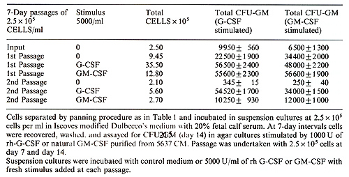
In order to substantiate the role of G-CSF as a true physiological
regulator of granulopoi esis, we investigated the action of rhG-CSF
in normal endotoxin-hyporesponsive C3H/ HeJ mice. The almost complete
biological and receptor cross-reactivity of normal and leukemic
hematopoietic cells to murine and human G-CSF [9] indicated that
the human recombinant molecule should have biological activity in
vivo in the murine system. Indirect evidence for this has been obtained
in clinical situations where tumor-associated neutrophil leucocytosis
could be transferred to nude mice bearing G-CSF-producing human
tumor implants [20-23]. Injection of rhG-CSF intravenously into
mice, with monitoring of serum levels by a human marrow or WEHI-3
leukemic colony assay, revealed a biphasic decay curve with a rapid
initial decline and a subsequent slower rate of decay with a half-life
of > 2.5 h. Intraperitoneal injections of 1.5 µg of rhG-CSF twice
daily produced serum levels of 5000075000 units/mI serum after 1
hand plateau levels were maintained for 6-8 h. Within 48 h of initiation
of daily G-CSF therapy, blood neutrophil numbers doubled and then
rose progressively to 26000/mmł by day 5 and fluctuated between
11 000 and 54000/ mmł thereafter. The average increase in blood
neutrophils was ninefold over control injected mice and the number
of blood monocytes, while variable, increased on average threefold
from days 5 to 14. The absolute number of eosinophils remained un
changed and a persisting mild lymphocytosis was induced with a twofold
increase from 4 to 14 days of treatment. The influence of G-CSF
therapy on hematopoietic tissue was particularly evident in the
spleen, whose cellularity increased threeto fourfold by day 7 of
G-CSF treatment with a conversion from a lymphoid tissue to a myeloid
organ with > 50% neutrophilic granulocytes at all stages of differentiation,
extensive erythropoiesis and marked megakaryocyte development (Fig.3).
Marrow cellularity was not changed following GCSF treatment but
the marrow population became almost exclusively granulopoietic with
loss of marrow lymphopoiesis and marked suppression of marrow erythropoietic
activity. Total neutrophil granulopoietic mass in treated animals
increased 4.5-fold by day 7 and the decrease in marrow erythropoiesis
was more than compensated for by an increase in splenic erythropoiesis,
with total erythroid mass more than doubling in treated mice. Megakaryocyte
numbers fell progressively in the marrow of treated mice, but the
marked increase in splenic megakaryocytes resulted in a threefold
increase in total body megakaryocytes. The increase in immature
erythroid and megakaryocytic cells was not associated with changes
in hematocrit or platelet levels. The total splenic lymphocyte population
remained unchanged and there was no evi dence of stress-induced
involution of thymic lymphopoiesis. Analysis of the various progenitor
populations responsive in colony assay to IL-3, CSF-l, GM-CSF, and
G-CSF revealed little change in the marrow compartment in GCSF-treated
mice, but a dramatic increase in the splenic CFU-GM population (50-
to 150-fold) resulted in an overall increase of IL-3-responsive
CFU-GM (fourfold) and of CSF-l-, GM-CSF-, and G-CSF-responsive CFU-GM
(threefold). Pluripotential stem cells detected in the day-12 CFU-S
assay showed that this compartment remained unchanged in the marrow,
but the marked increase in splenic CFU-S resulted in a 2.5fold total
body increase in these stem cells. 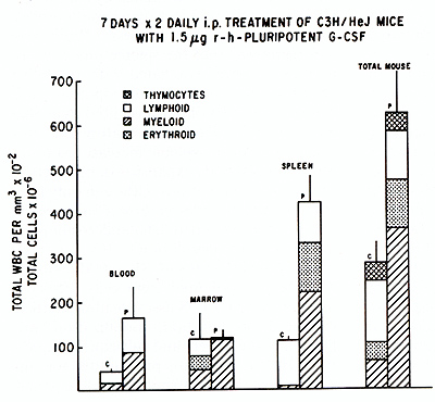
In vitro studies using accessory cell-depleted marrow indicate that the direct action of hG-CSF is restricted primarily to proliferation and differentiation in the neutrophil granulocyte lineage [14] and the factor does not support directly differentiation into the erythroid, megakaryocytic or eosinophillineage. Evidence for a possible direct action on early stem-cell proliferation and subsequent generation of BFU- E, CFU -G EMM, and megakaryocyte (CFU-M) is equivocal [15] and awaits a demonstration of G-CSF receptors on enriched populations of these cells or a proliferative response involving pure populations of such cells. Here we have documented that G-CSF can stimulate the generation of new CFU -G M in suspension culture of accessory cell-depleted human marrow, suggesting a recruitment action on some pre-CFU-GM population and/or enhanced self-renewal of preexisting CFUGM, capable of responding to both G-CSF and GM-CSF. This observation provides evidence for a stimulatory action of G-CSF on a progenitor population with a differentiation potential more extensive than that of neutrophil lineage-restricted CFU-GM, since the CFU-GM generated in suspension cultures with G-CSF responded to GM-CSF in colony assay by a normal pattern of differentiation into eosinophils and macrophages as well as neutrophils. In vivo administration of G-CSF by the intraperitoneal route led to sustained serum levels of bioactive G-CSF comparable to those found 3-6 h post injection of endotoxin in endotoxin-responsive strains of mice [6]. The use of highly purified recombinant G-CSF, the absence of significant contamination of our G-CSF with endotoxin, and the use of C3H/HeJ mice allow us to conclude that the in vivo responses seen were due to the action of G-CSF , rather than any contaminant acting directly or synergistically on neutrophil production. The predominant response involved elevation of peripheral blood neutrophils by a factor of 6-9 in C3H/HeJ mice, which was somewhat lower than we have observed in other strains, since up to a 20-fold increase in blood neutrophils can be obtained in Balb/c mice. Studies in hamsters also indicated a specific action on the neutrophil lineage with a three- to sixfold increase in peripheral blood neutrophils [24], and in monkeys receiving 10µg rhG-CSF per kilogram per day, neutrophil counts rose fivefold, and up to 12-fold with lOO µg/kg per day [25]. In mice, demand for increased hematopoiesis following such insults as endotoxin treatment, irradiation, or cytotoxic drug treatment is met by a marked expansion of splenic hematopoiesis with little absolute change in marrow cellularity. Thus, it was not surprising that the major expansion of neutrophil production elicited by chronic GCSF treatment was met by a rapid onset of extensive splenic myelopoiesis. What was surprising was that the splenic phase of hematopoiesis was associated with increased erythropoiesis, megakaryocytopoiesis, and elevated numbers of pluripotential stem cells and progenitors of all hematopoietic lineages. Indeed, the splenic increase led to an absolute total body expansion of production of these cell populations, although only neutrophils and to a lesser extent monocytes, and not red cells, platelets, and eosinophils increased in the circulation. It is possible that G-CSF acts directly to activate pluripotential stem cells, detected by the day-12 CFU-S assay, leading to their enhanced proliferation, mobilization, migration to the spleen, and subsequent expansion. In this scenario, progenitors for lineages other than neutrophil could be gener ated in increased numbers as a result of stochastic mechanisms believed to operate at the level of pluripotential stem-cell self-renewal and differentiation. Alternatively, indirect mechanisms may operate, whereby G-CSF-induced differentiation depletes a population of neutrophil progenitors, which in turn triggers activation of the pluripotential stem-cell pool and expansion of the marrow microenvironment. A third possibility is that G-CSF acts in vivo upon an accessorycell population to induce release of other hematopoietic growth factors known to influence multiple hematopoietic lineages. This third possibility was suggested by the original observations that G-CSF stimulated BFU-E and CFU-GEMM in marrow cultures depleted of T cells but not otherwise depleted of accessory cells. Indirect induction of a growth factor acting on pluripotential stem cells has been invoked as a mechanism to explain the marked stimulation of splenic hematopoiesis following administration of endotoxin [26]. Staber et al. [26] reported a mouse serum factor induced following endotoxin treatment, that, when injected into normal mice, elicited splenomegaly and a marked rise in splenic CFU-GM by 5 days, with concordant elevation of erythroid and megakaryocytic progenitors. The serum factor was thought not to be G M CSF or residual endotoxin, although its peak appearance in the serum between 30 min and 3 h after endotoxin injection would coincide with the kinetics of endotoxin-induced G-CSF. This serum factor stimulating murine splenic hematopoiesis could be G-CSF itself or a molecule induced by G-CSF and mediating the more general effects on stem-cell proliferation and mobilization seen in the in vivo studies. The stimulation of increased granulopoiesis persists as long as G-CSF is administered in vivo, although the duration of studies to date has been limited to 4-5 weeks. In longterm marrow culture much longer periods, up to 4 months with sustained high levels of G-CSF, have been obtained with persisting elevation of neutrophil production relative to control cultures, and no evidence was obtained for premature exhaustion of stem cells or accelerated depletion of cultures. The ability of low concentrations of TNF alpha to counteract the proliferative action of G-CSF in vitro in clonal assay and longterm bone marrow cultures suggests a physiological role for TNF as a natural inhibitor of neutrophil granulopoiesis. The coordinate inducibility of G-CSF and TNF alpha following endotoxin activation of macrophages may provide a mechanism self-limiting the chronic generation of neutrophils from marrow progenitors following bacterial infection. Alternatively, the relative resistance to TNF inhibition of myeloid progenitors stimulated by GM-CSF may provide an alternate pathway for the generation of neutrophils in the face of elevated TNF levels. The interrelationship of TNF and CSF in the production and function of neutrophils is further emphasized by the observation that TNF can enhance both the phagocytic ability and the antibody-dependent cytotoxicity of neutrophils, increase their super oxide anion production, and stimulate their adherence to endothelial cells [27]. In addition, TNF has been shown to stimulate growth factor production by endothelial cells, due in part to activation oftranscription of the GM-CSF gene [28]. The interplay of direct marrow-suppressive action of TNF and its role in mature neutrophil activation and induction of CSF production by accessory cells such as endothelium must be considered when extrapolating to an in vivo action of TNF in granulopoiesis.
1. MetcalfD, Nicola NA (1983) Proliferative effects of purified granulocyte colony-stimulating factor (O-CSF) on normal hemopoietic cells. J Cell Physiol116: 198 2. Platzer E, Welte K, Oabrilove J, Lu L, Harris P, Mertelsmann R, Moore MAS (1985) Biological activities of a human pluripotent hemopoietic colony-stimulating factor on normal and leukemic cells. J Exp Med 162: 1788 3. Metcalf D (1986) Review: the molecular biology and function of the granulocyte-macrophage colony-stimulating factors. Blood 67:257 4. Nicola NA, Metcalf D, Matsumoto M, Johnson OR (1983) Purification of a factor inducing differentiation in murine myelomonocytic leukemia cells: identification as granulocyte colony-stimulating factor (O-CSF). J Biol Chem 258:9017 5. Burgess A W , M etcalf D ( 1980 ) Characteriza tion of a serum factor stimulating the differentiation of myelomonocytic leukemic cells. Int J Cancer 26:647 6. Moore MAS (1982) G-CSF: its relationship to leukemia differentiation-inducing activity and other hemopoietic regulators. J Cell Physiol [Suppl] 1 :53 7. Tsuda H, Neckers LM, Pluznik EH (1986) Colony-stimulating factor-induced differentiation of murine M 1 myeloid leukemia cells is permissive in early Gl phase. Proc Natl Acad Sci USA 83:4317 8. Nicola NA, MetcalfD, Johnson GR, Burgess A W ( 1979) Separation of functionally distinct human granulocyte-macrophage colonystimulating factors. Blood 54:614 9. Nicola NA, Begley CG, Metcalf D (1985) Identification of the human analogue of a regulator that induces differentiation in murine leukaemic cells. Nature 314:625 10. Welte K, Platzer L, Lu L, Gabrilove JL, Levi E, Mertelsmann R, Moore MAS (1985) Purification and biochemical characterization of human pluripotent hematopoietic colonystimulating factor. Proc Natl Acad Sci USA 82:1526 11. Gabrilove JL, Welte K, Harris P, Platzer E, Lu L, Levi E, Mertelsmann R, Moore MAS (1986) Pluripoietin alpha: a second human hematopoietic colony-stimulating factor produced by the human bladder carcinoma cell line 5637. Proc Natl Acad Sci USA 83:2478 12. Souza LM, Boone TC, Gabrilove J, Lai PH, Zsebo KM, Murdock DC, Chazin VR, Bruszewski J, Lu H, Chen KK, Barendt J, Platzer E, Moore MAS, Mertelsmann R, Welte K (1986) Recombinant human granulocyte colony-stimulating factor: effects on normal and leukemic myeloid cells. Science 232:61 13. Tsuchiya M, Asano S, Kaziro Y, Nagata S (1986) Isolation and characterization of the cDNA for murine granulocyte colony-stimulating factor. Proc Natl Acad Sci USA 83:7633 14. Ottmann OG, Welte K, Souza LM, Moore MAS (in press) Proliferative effects of a recombinant human granulocyte-colony stimulating factor (rG-CSF) on highly enriched hematopoietic progenitor cells. This volume 15. MetcalfD, Nicola NA (1983) Proliferative effects of purified granulocyte colony-stimulating factor (G-CSF) on normal mouse hemopoietic cells. J Cell Physiol116: 198 16. Vadas MA, Lopez AF (1985) Regulation of granulocyte function by colony-stimulating factors and monoclonal antibodies. Lymphokines 12:179 17. Shah RG, Green S, Moore MAS (1978) Colony-stimulating and inhibiting activities in mouse serum after c. parvum-endotoxin treatment. J Reticuloendothel Soc 23:29 18. Aggarwal BB, Kohr WJ, Hass PE, Moffat B, Spencer SA, Henzel WJ, Bringman TS, Nedwin GE, Goeddel DV, Harkins RN (1985) Human tumor necrosis factor: production, purification and characterization. J BioI Chem 260:2345 19. Broxmeyer HE, Williams DE, Lu L, Cooper S, Anderson SL, Beyer GS, Hoffman R, Rubin BY (1986) The suppressive influence of human tumor necrosis factors on bone marrow hematopoietic progenitor cells from normal donors and patients with leukemia: synergism of tumor necrosis factor and interferon-gamma. J Immuno1136:4487 20. Asano S, Urabe A, Okabe T, Sato N, Kondo Y, Ueyama Y, Chiba S, Ohsawa S, Kosaka K (1977) Demonstration of granulopoietic factor(s) in the plasma of nude mice transplanted with human lung cancer and in tumor tissue. Blood 49:845 21. Sato N (1979) Granulocytosis and colonystimulating activity (CSA) by a human squamous cell carcinoma. Cancer 43:605 22. Mizoguchi H, Suda T, Miura Y, Kubota K, Takaku F (1982) Hemopoietic stem cells in nude mice transplanted with colony-stimulating-factor-producing tumors. Exp Hemat 10:874 23. Motoyoshi K, Suda T, Takaku F, Miura y (1983) Regulatory mechanism of granulopoiesis in the bone marrow ofCSF-producing tumor-bearing nude mice. Blood 62:980 24. Zsebo KM, Cohen AM, Murdock DC, Boone TC, Inoue H, Chazin VR, Hines D, Souza LM (1987) Recombinant human granulocyte colony-stimulating factor: molecular and biological characterization. Immunobiology (in press ) 25. Welte K, Bonilla MA, Gillio AP, Boone TC, Potter GK, Gabrilove JL, Moore MAS, O'Reilly RJ, Souza LM (1987) Recombinant human G-CSF: effects on hematopoiesis in normal and cyclophosphamide-treated primates. J Exp Med (in press) 26. Staber FG, Burgess A W , Nicola NA, Metcalf D (1984) Biological and biochemical properties of a serum factor that stimulates splenic hemopoiesis in mice. Exp Hematol12: 107 27. Shalaby MR, Pennica D, Palladino MA Jr ( 1986) An overview of the history and biological properties of tumor necrosis factor. Springer Semin Immunopathol 9:33 28. Broudy VC, Kaushansky K, Segal GM, Harlan JM, Adamson JW (1986) Tumor necrosis factor type alpha stimulates human endothelial cells to produce granulocyte/macrophage colony-stimulating factor. Proc Natl Acad Sci USA 83:7467 |