|
Department of Biochemistry and Laboratory of Molecular Biology, Shanghai Second Medical University, Shanghai, People's Republic of China Alterations of cellular oncogene expression have been reported in numerous neoplasias, particularly of the hematopoietic system, However, a cause-effect relationship between cellular oncogene abnormalitics and oncogenesis is still unclear, In order to explore the involvement of cellular oncogenes in human cancer cells, we studied the levels of the expression of three cellular protooncogenes in the primary leukemic cells from 53 leukemic patients with different types and stages.
Thirty-five patients with acute myeloblastic Icukemia (AML), 14
with acute lymphoblastic leukemia (ALL), 2 with chronic myelocytic
leukemia (CML), 2 with chronic lymphocytic leukemia (CLL), and 8
normal individuals as controls were studied. Diagnosis was based
on the morphological evaluation of bone marrow smears according
to the FrenchAmerican-British (FAB) criteria. Leukemic cells, obtained
from peripheral blood after diagnosis and before initiation of chemotherapy,
were enriched up to 90% by centrifugation on Ficoll-Hypaque density
gradient [1]. HL-60, a human promyelocytic leukemia cell line, was
used as reference.
I. Storage of Samples 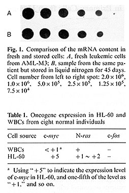 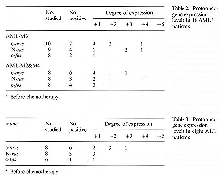 II, Expression Levels of Protooncogenes in Normal Leukocytes The expression levels of c-myc, N-ras, and c-fos in peripheral leukocytes from eight different normal individuals, and the HL-60 cell line were detected. The level of the c-myc transcripts in HL-60 was denoted as ..+ 5" [3], and the onefifth of the level as ..+ 1" and so on. Figure 2 and Table 1 show that the myc gene was slightly expressed and N-ras was marginally expressed, whereas the expression of c-fos was undetectable in the normal leukocytes. III. Expression Levels of Protooncogenes in Leukemic Cells In ten AML-M3, four AML-M2, four AML-M4, and ten ALL patients, before chemotherapy, the c-myc was obviously expressed in almost all leukemic cells irrespective of the cell types, while N-ras and c-fos were unconstantly expressed. However, the C-fos was expressed in all four cases ofAML-M4 (Tables 2,3). The c-myc transcripts were detected but the N-ras and c-fos were not in four chronic leukemic cases. The samples were obtained from the three AML patients 2- 3 months after initial therapy, while the patients were in remission and on maintenance chemotherapy. The results showed that the levels ofc-myc mRNA decreased obviously in one of three remission samples (Fig. 3), while the levels of c-myc mRNA remained essentially unchanged in the others. Figure 4 demonstrates that c-myc and N-ras mRNA increased dramatically in CLL during blast crisis. 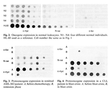
In this experiment, we selected three cellular protooncogenes -c-myc and c-fos gene coding for nuclear proteins, the former relating to early cell differentiation and the latter linking to monocytic differentiation and N-ras for putative intermediate transducers of mitogenic signals for detecting their expression levels in primary cells from human leukemia. Most previous studies on the expression of cellular oncogenes in leukemias have been carried out on neoplastic lines, which may not faithfully represent the primary cancer cells [ 4- 6] .So in this study we observed the expression of protooncogenes in primary cells from human leukemias. Our results showed that only c-myc gene expressed slightly in normal leukocytes while expressed obviously in almost all leukemic cells irrespective of the cell types. The c-myc expression level is higher in acute leukemia than in chronic leukemia. But the levels of N-ras and c-fos transcripts were variable (Tables 2, 3). An interesting finding is that the c-fos gene was expressed in all four AML-M4. The recent data indicate that the N-ras gene may be activated by mutation in AML [7]. Our results showed the N -ras was strictly expressed in acute leukemia, but there is no correlation between the quantitative expression in AML and ALL. So the level of N-ras expression may not be significantly changed in leukemic cells. Until now there have only been a few articles written about oncogene expression during remission and after chemotherapy [8]; we analyzed three cases of AML and one of CLL, but no definite suggestion can be put forward. We think that it is important to study the changes of oncogene expression in the course of remission.
1. Ferrante A, Thong YH (1978) A rapid one step procedure for purification
of mononuclear and polymorphonuclear leukocytes from human blood
using a modification of the Hypaque-Ficoll technique. J Immunol
Methods 24: 389- 393 |